Use of imageJ software to evaluate the radiological repair of circular bone defects treated with demineralized bone matrix in rabbits
DOI:
https://doi.org/10.19137/cienvet202224204Keywords:
Radiodensity, ImageJ, Bon defect, Demineralized bone defect, RabbitAbstract
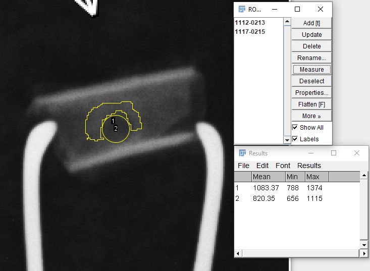
Radiology is a technique used in the diagnosis of fractures and evaluation of bone repair processes. The interaction of the X-rays with the tissues produces a gray image whose density can be set with ImageJ software. The authors report the use of the software to assess the repair progress of orthopedic bone defects treated with demineralized bone matrix (DBM). 12 rabbits that formed 3 treatment groups were used. The animals had a circular defect created in one of the femoral shafts that were treated with MOD. The samples were obtained at 15, 30 and 60 days postoperatively and X-rays of the defects were taken, quantifying the densities of the defects and the neighboring bone; the ratio between both densities generated the relative optical density (DOR) of the defect. The DORs were analyzed in the Infostat software. Radiographs indicated increased densities of the defects, consistent with repair. At 15 days, the mean DOR was 0.68 (±0.08); at 30 days 0.7 (±0.08), and at 60 days 0.94 (±0.03). Fisher’s LSD test established that there were no significant differences at 15 and 30 days, while at 60 days the increase in densities was significant (p>0.05). The method used to analyze radiological images with ImageJ validates the comparison of densities at different stages of repair of MOD-treated orthopedic bone defects.
Downloads
References
2. Bigham-Sadegh A, Karimi I, Alebouye M, Shafie-Sarvestani Z, Oryan A. Evaluation of bone healing in canine tibial defects filled with cortical autograft, commercial-DBM, calf fetal DBM, omentum and omentum-calf fetal DBM. J. Vet. Med 2013; 14(3), 337. doi:10.4142/jvs.2013.14.3.337
3. Bohner M, Miron RJ. A proposed mechanism for material-induced heterotopic ossification. Materials Today. 2019; 22:132–141. DOI: 10.1016/j.mattod.2018.10.036.
4. Audisio SA, Vaquero PG, Torres PA, Verna EC, Ocampo LN, Ratusnu V, et al. Obtención – caracterización y almacenamiento de matriz ósea desmineralizada. Revista de Medicina Veterinaria 2015; 17 (2):24-34.
5. Mol A. Instrumentos de procesamiento de imagen para aplicaciones dentales. Miles Dale A: Clínicas Odontológicas Norteamericanas. Vol. 2 Aplicaciones de las distintas modalidades de la imagen digital en odontología. Mexico 2000. McGraw- Hill Interamericana, 482p.
6. Chiapasco M, Rossi A, Motta JJ, Crescentini M. Spontaneous bone regeneration after enucleation of large mandibular cysts: a radiographic computed analysis of 27 consecutive cases. J. Oral Maxillofac 2000; 58(9), 942–948. doi:10.1053/joms.2000.8732
7. Di Rienzo JA, Casanoves F, Balzarini MG, Gonzalez L, Tablada M, Robledo CW. InfoStat versión 2016. [Internet] Grupo InfoStat, FCA, Universidad Nacional de Córdoba, Argentina. URL http://www.infostat.com.ar
8. Ihan Hren N, Miljavec M. Spontaneous bone healing of the large bone defects in the mandible Int. J. Oral Maxillofac. Surg. 2008; 37(12):1111-1116. doi: 10.1016/j.ijom.2008.07.008
9. Schindelin J, Arganda-Carreras I, Frise E, Kaynig V, Longair M, Pietzsch T, et al. Fiji: an open-source platform for biological-image analysis. Nat Methods 2012 28; 9 (7):676-82. doi: 10.1038/nmeth.2019
Downloads
Published
How to Cite
Issue
Section
License
Al momento de enviar sus contribuciones, los colaboradores deberán declarar , de manera fehaciente, que poseen el permiso del archivo o repositorio donde se obtuvieron los documentos que se anexan al trabajo, cualquiera sea su formato (manuscritos inéditos, imágenes, archivos audiovisuales, etc.), permiso que los autoriza a publicarlos y reproducirlos, liberando a la revista y sus editores de toda responsabilidad o reclamo de terceros , los autores deben adherir a la licencia Creative Commons denominada “Atribución - No Comercial CC BY-NC-SA”, mediante la cual el autor permite copiar, reproducir, distribuir, comunicar públicamente la obra y generar obras derivadas, siempre y cuando se cite y reconozca al autor original. No se permite, sin embargo, utilizar la obra con fines comerciales.


.jpg)

4.png)