Pathological and Echocardiographic Key Points of Right Atrioventricular Valve Dysplasia
DOI:
https://doi.org/10.19137/cienvet202325104Keywords:
Dysplasia, Tricuspid, Canine, Feline, Echocardiography, PathologyAbstract
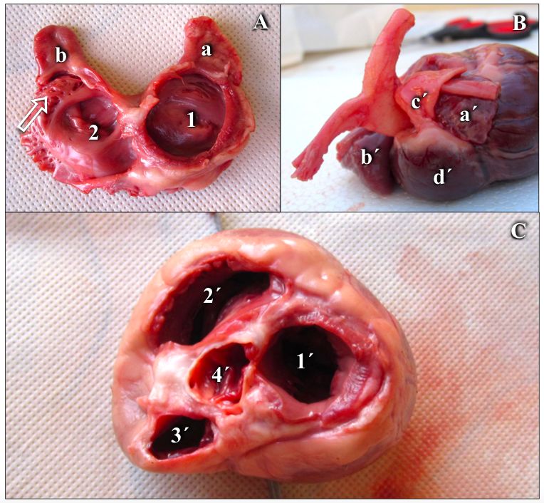
Congenital insufficiency of the right atrioventricular valve is defined as tricuspid dysplasia. The alteration occurs in septal and mural valve leaflets, in chordae tendineae or in papillary muscles. The malformation causes tricuspid regurgitation and presentation of clinical signs associated with congestive right heart failure. Macroscopic lesions include localized or diffuse nodulation or thickening of valves leaflets, short or elongated chordae tendineae, with incomplete development and partial fusion or union, papillary muscles with abnormal attachment or incomplete separation to the right ventricular wall, or absence of one or more muscles. The aim of this work is to provide information and casuistry for diagnostic purposes of a pathology of low prevalence in companion animals. Between 2011 and 2021, 24 canines with a definitive diagnosis of tricuspid dysplasia were treated. The pathology was more frequent in dogs of small to medium size, Poodle (4), Dachshund (3), French Bulldog (3), Yorkshire terrier (2), Bull terrier (1), Boxer (1) and 10 mongrels, 3 under 12 kg and 7 between 12 and 30 kg in weight, medium to large size. The findings are different, race and size, to those observed by other authors. Nodular thickening associated with short chordae tendineae was the most common valve malformation. Tricuspid dysplasia was associated with half (13 of 24) of the cases with pulmonary stenosis. Other congenital anomalies, interventricular and atrial septal defect, were also related. The precise diagnosis of the congenital abnormality of the tricuspid valve apparatus can only be made by echocardiography. Valve alterations, septal leaflet, characterized by nodulation, thickening and union to the
interventricular septum, were the most common. The continuous spectral Doppler exploration allowed determining severity, pressures in the atrial and right ventricular chambers and establishing hemodynamic effects of the process and its implication in the prognosis and treatment of the patient.
Downloads
References
2. Takemura N, Machida N, Nakagawa K. et al. Ebstein´s anormaly in a beagle dog. Journal Veterinary Medicine Science.2003; 65:531-533.
3. Dyce KM, Sack WO, Wensing CJG. Anatomía Veterinaria. 2º Edición. McGraw-Hill Interamericana. Philadelphia.1996. 7:235-251
4. Boon JA. Shunts congénitos y displasia de la válvula auriculoventricular. En: Boon, J.A. Ecocardiografía Veterinaria. 2º Edición. Multimédica
Ediciones Veterinarias.2012. Barcelona-España.9:313-339.
5. Armstrong WF, Ryan T. Ecocardiografía de Feigenbaum. 7º Edición. Lippincott Williams & Wilkins. 2011. ISBN 978-84-96921-82-5. 13:337-
359.
6. Becker AE, Becker MJ. and Edwards JE. Pathologic Spectrum and Dysplasia of the Tricuspid Valve: Features in Common with Ebstein’s Malformation. Archives of Patholoy.1971; 91,167-178
7. Oyama MA, Sisson DD, Thomas WP, Bonagura JD. Cardiopatías Congénitas. En: Ettinger, S.J.; Feldman, E.C. Tratado de Medicina Interna Veterinaria. 2007. 6º Edición. 2(200):972-1022.
8. Moise NS. Tricuspid valve dysplasia in the dog. In:Current Veterinary Therapy XII. Eds WB Saunders, Kirk RW, Bonagura JD,
1995.Philadelphia. pp.813–816
9. Liu SK, Tilley LP. Dysplasia of the tricuspid valve in the dog and cat. Journal of American Veterinary Medical Association. 1976; 169(6):623-
630.
10. Kittlesson MD, Kienle RD. Anomalías congénitas de las válvulas auriculoventriculares. En: Medicina cardiovascular de pequeños animales. 2000. Multimédica. Barcelona. 17:273-281.
11. Ware WA. Cardiopatías congénitas comunes. En: Nelson RW, Couto CG. Medicina Interna de Animales Pequeños. 1(9):161-179. Wright NK,
Bleas ME, Benson DW. (2001). Clinical spectrum of congenital tricuspid valve malformations in an extended family of Labrador Retrievers. In: Proceedings of the 19th ACVIM Congress, 2005.Denver, USA, 2001:95
12. Strikland KN. Cardiopatía Congénita. En: Manual de Cardiología Canina y Felina. 4º Edición. Multimédica Ediciones Veterinarias. 2009. Barcelona.
2(12):189-209.
13. Siemens M, Davidson AP. Tricuspid valve dysplasia in Labrador Retrievers. In: Proceedings of the 19th ACVIM Congress, Denver, 2001. USA, 2001:95.
14. Hoffmann G, Amberger CN, Seiler G. et al. Tricuspid valve dysplasia in fifteen dogs. Schweiz Archive Tierheilkd.2000. 142:268-277.
15. Chetboul V, Tran D, Carlos C. et al. Congenital malformations of the tricuspid valve in domestic carnivores: a retrospective study of 50 cases. 2004. Schweiz Archive Tierheilkd. 146:265-275.
16. Sousa MG, Gerardi1 DG, Alves RO, Camacho AA. Tricuspid valve dysplasia and Ebstein’s anomaly in dogs: case report. Arq. Bras. Med. Vet.
Zootec.2006; 58(5):762-767.
17. Paslawska U, Noszczyk-Nowak A, Janiszewski A, Nicpón J. Tricuspid dysplasia in dogs. Bull Vet Inst Pulawy. 2013; 57:123-126. DOI: 10.2478/ bvip-2013-0023.
18. Navarro-Cubas X, Palermo V, French A, Sanchis-Mora S, Culshaw G. Tricuspid valve dysplasia: A retrospective study of clinical features and outcome in dogs in the UK. Open Veterinary Journal.2017; 7(4): 349- 359. DOI: http://dx.doi.org/10.4314/ovj.v7i4.11
19. Favril S, Broeckx BJG, de Rooster H. et al. Tricuspid valve dysplasia in dogs. Vlaams Diergeneeskundig Tijdschrift.2018; 87:14-21.
20. Kaplan, P.M. (1991). Congenital heart disease. Problem Veterinary Medicine. 3:500-519.
21. K Bonagura JD, Luis Fuentes V. Ecocardiografía. En: Mattoon, J.S.; Nyland, T.G. Diagnóstico Ecográfico en Pequeños Animales.2016. 3º Edición. Multimédica Ediciones Veterinarias. Barcelona. 8:245-383
Downloads
Published
How to Cite
Issue
Section
License
Al momento de enviar sus contribuciones, los colaboradores deberán declarar , de manera fehaciente, que poseen el permiso del archivo o repositorio donde se obtuvieron los documentos que se anexan al trabajo, cualquiera sea su formato (manuscritos inéditos, imágenes, archivos audiovisuales, etc.), permiso que los autoriza a publicarlos y reproducirlos, liberando a la revista y sus editores de toda responsabilidad o reclamo de terceros , los autores deben adherir a la licencia Creative Commons denominada “Atribución - No Comercial CC BY-NC-SA”, mediante la cual el autor permite copiar, reproducir, distribuir, comunicar públicamente la obra y generar obras derivadas, siempre y cuando se cite y reconozca al autor original. No se permite, sin embargo, utilizar la obra con fines comerciales.


4.png)