Quantitative histopathological analysis of a canine squamous cell carcinoma using digital image processing and machine learning-based classification
Quantitative histopathological analysis of a canine squamous cell carcinoma using digital image processing and machine learning-based classification
DOI:
https://doi.org/10.19137/cienvet.v28.10055Keywords:
Carcinoma de células escamosas, Patologia digital, Machine learning, Procesamiento de imágenes, Random Forest, PerroAbstract
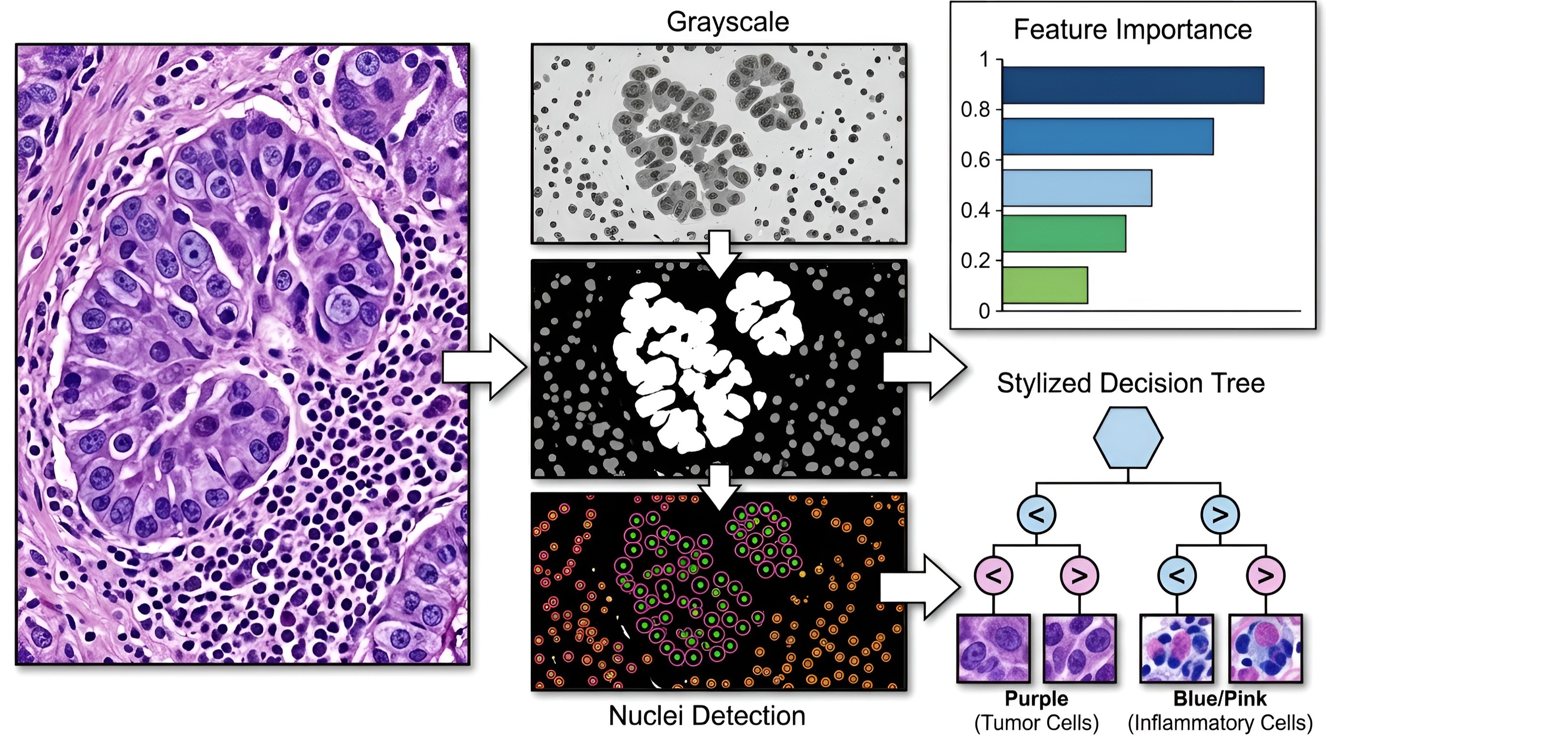
Squamous cell carcinoma (SCC) is one of the most frequent malignant cutaneous neoplasms in dogs, and its diagnosis relies on the subjective histopathological evaluation by the pathologist. The objective of this study was to develop and evaluate a computational pipeline for quantitative analysis of histopathological images of canine SCC, using digital image processing and machine learning-based cell classification. A digital photomicrograph of a canine cutaneous SCC (H&E stain) was processed using Otsu thresholding, morphometric filtering, and extraction of cellular descriptors (area, perimeter, eccentricity, solidity, and circularity). Seventy-nine regions of interest were identified, of which 63 were manually classified by an expert pathologist (33 tumorous, 30 inflammatory). A Random Forest classifier (100 trees) was trained with a 70/30 stratified split. The model achieved 52.6% accuracy (macro F1 = 0.52) in the test set, with circularity as the most important descriptor (relative importance: 0.229). Tumor cells showed larger area (1227 ± 876 px) and eccentricity (0.73 ± 0.17) than inflammatory cells (1034 ± 779 px; 0.69 ± 0.14). The results demonstrate that traditional morphometric features allow preliminary differentiation between cell populations, but their discriminatory power is limited, warranting deep learning approaches in future studies. The implemented pipeline constitutes an accessible and reproducible tool for objective quantification in veterinary pathology
References
1. Withrow SJ, Vail DM, Page RL. Withrow & MacEwen's small animal clinical oncology. 5th ed. St. Louis: Elsevier Saunders; 2013.
2. Goldschmidt MH, Goldschmidt KH. Classification of epithelial and melanocytic tumors of the skin of dogs and cats. In: Meuten DJ, editor. Tumors in domestic animals. 5th ed. Ames: Wiley-Blackwell; 2017. p. 164-169.
3. Gross TL, Ihrke PJ, Walder EJ, Affolter VK. Skin diseases of the dog and cat: clinical and histopathologic diagnosis. 2nd ed. Oxford: Blackwell Science; 2005.
4. Peiffer RL, Spencer C, Popp JA. Grades of malignancy and prognosis in canine cutaneous squamous cell carcinoma. Vet Pathol. 1984;21(1):95-96. Disponible en: https://doi.org/10.1177/030098588402100118
5. van der Laak J, Litjens G, Ciompi F. Deep learning in histopathology: the path to the clinic. Hum Pathol. 2021; 110:11-22. Disponible en: https://doi.org/10.1016/j.humpath.2020.11.007
6. Otsu N. A threshold selection method from gray-level histograms. IEEE Trans Syst Man Cybern. 1979;9(1):62-66. Disponible en: https://doi.org/10.1109/TSMC.1979.4310076
7. Breiman L. Random forests. Mach Learn. 2001;45(1):5-32. Disponible en: https://doi.org/10.1023/A:1010933404324
8. van der Walt S, Schönberger JL, Nunez-Iglesias J, Boulogne F, Warner JD, Yager N, et al. scikit-image: image processing in Python. PeerJ. 2014;2: e453. Disponible en: https://doi.org/10.7717/peerj.453
9. Pedregosa F, Varoquaux G, Gramfort A, Michel V, Thirion B, Grisel O, et al. Scikit-learn: machine learning in Python. J Mach Learn Res. 2011; 12:2825-2830.
10. Gonzalez RC, Woods RE. Digital image processing. 4th ed. New York: Pearson; 2018.
11. Meuten DJ, editor. Tumors in domestic animals. 5th ed. Ames: Wiley-Blackwell; 2017.
12. Meuten DJ, Everitt JF, Reindel JH, Moore RR. Canine cutaneous squamous cell carcinoma: a review of 55 cases. Vet Pathol. 1985;22(5):485. Disponible en: https://doi.org/10.1177/030098588502200515
13. Haralick RM, Shanmugam K, Dinstein I. Textural features for image classification. IEEE Trans Syst Man Cybern. 1973; SMC-3(6):610-621. Disponible en: https://doi.org/10.1109/TSMC.1973.4309314
14. Couteaux V, Si-Mohamed S, Nempont O, Leforestier G, Salomon A, Pellegrinelli J, et al. Deep learning-based classification of metastatic breast cancer histopathology images. Proc IEEE Int Symp Biomed Imaging. 2019; 2019:1110-1113. Disponible en: https://doi.org/10.1109/ISBI.2019.8759482
15. Echle A, Rindtorff NT, Brinker TJ, Luedde T, Pearson AT, Kather JN. Deep learning in cancer pathology: a new generation of clinical biomarkers. Br J Cancer. 2021;124(4):686-696. Disponible en: https://doi.org/10.1038/s41416-020-01122-x
Published
Issue
Section
License
Copyright (c) 2026 Arnulfo Villanueva Castillo, César F. Pastelín-Rojas, Hermilo Lucio-Castillo, Jesús J. Hinojosa-Moya, Fabiola Rodríguez-Andrade, Miguel Á. Zambrano-González, Erick C. Fernández-Meneses, Briseida L. Castro-Bautista, Claudia Mancilla-Simbro, Ruby Sandy Moreno-Mejía, Alberto Ramírez-Mata

This work is licensed under a Creative Commons Attribution-NonCommercial-ShareAlike 4.0 International License.
Al momento de enviar sus contribuciones, los colaboradores deberán declarar , de manera fehaciente, que poseen el permiso del archivo o repositorio donde se obtuvieron los documentos que se anexan al trabajo, cualquiera sea su formato (manuscritos inéditos, imágenes, archivos audiovisuales, etc.), permiso que los autoriza a publicarlos y reproducirlos, liberando a la revista y sus editores de toda responsabilidad o reclamo de terceros , los autores deben adherir a la licencia Creative Commons denominada “Atribución - No Comercial CC BY-NC-SA”, mediante la cual el autor permite copiar, reproducir, distribuir, comunicar públicamente la obra y generar obras derivadas, siempre y cuando se cite y reconozca al autor original. No se permite, sin embargo, utilizar la obra con fines comerciales.
How to Cite





.jpg)