Correlación entre los hallazgos imagenológicos y las características histopatológicas de adenocarcinomas de glándulas mamarias caninas: estudio de un caso clínico
Correlation between imaging findings and histopathological characteristics of canine mammary gland adenocarcinomas: clinical case study
DOI:
https://doi.org/10.19137/cienvet.v28.9575Palabras clave:
Neoplasia, Tumores caninos, Rayos x, Ecografía, HistologíaResumen
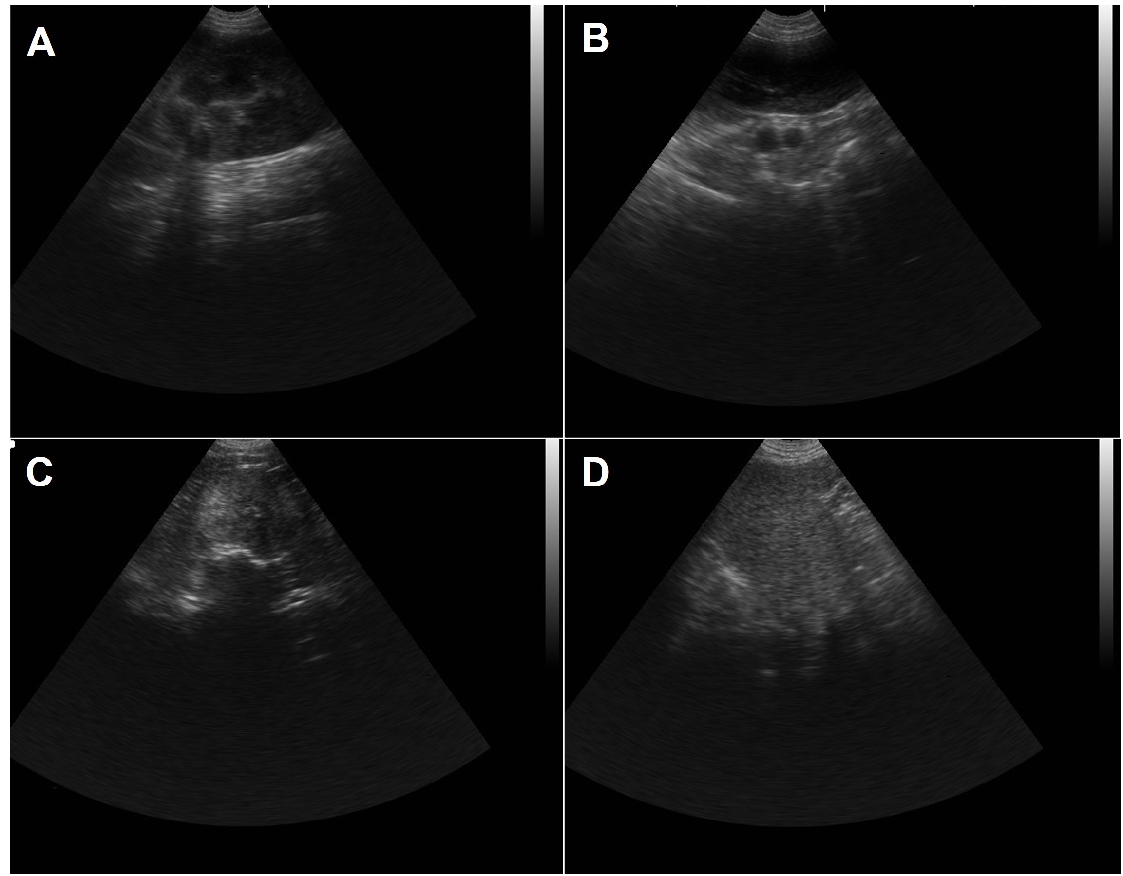
Los adenocarcinomas mamarios caninos constituyen la neoplasia más frecuente, con un 50% de casos malignos. Aunque la escisión quirúrgica es el tratamiento principal y la quimioterapia adyuvante mejora la supervivencia, el pronóstico suele ser desfavorable, con alta tasa de metástasis a ganglios linfáticos y pulmones. Este estudio correlaciona hallazgos obtenidos por medio de técnicas de imagenología con análisis histopatológico en un caso clínico de un can femenino Husky Siberiana de 10 años de edad. Las radiografías descartaron metástasis a distancia. La ecografía mamaria reveló masas heterogéneas, de bordes irregulares y mal definidos, con áreas hipoecoicas e hiperecoicas, vascularización aumentada, microcalcificaciones y áreas anecoicas por necrosis. Este patrón ecográfico fue altamente sugestivo de malignidad, lo que coincidió con el análisis histopatológico que confirmó la presencia de un carcinoma de tipo complejo, una neoplasia epitelial maligna caracterizada por dos poblaciones celulares. Las características ecográficas se correlacionaron directamente con la histología, las áreas hipoecoicas con el componente epitelial maligno y la necrosis, y las hiperecoicas con el estroma fibroso benigno, lo que nos permite concluir que la ecografía, una herramienta de rutina accesible, puede proporcionar un diagnóstico presuntivo altamente confiable de adenocarcinoma mamario maligno. El patrón descrito justificaría la recomendación de una extirpación quirúrgica total, pudiendo la biopsia histológica reservarse para la confirmación definitiva y la determinación del grado tumoral, crucial para el pronóstico y el tratamiento posterior. Esta correlación mejora el diagnóstico y contribuye a la oncología comparativa, que aprueba el uso de modelos caninos para el estudio del cáncer de mama
Referencias
Biondi L, Vannucci M, Boffoni L, Pereira P, Zaidan M. Quantification of Global DNA Methylation in Canine Mammary Gland Tumors via Immunostaining of 5-Methylcytosine: Histopathological and Clinical Correlations. Front Vet Sci. 2021;8:628241. DOI: 10.3389/fvets.2021.628241.
Kawamura Y, Itou H, Kida A, Sunakawa H, Kawamura K. Microwave ablation for the control of bleeding from disintegrated mammary tumours in two dogs. Vet Med Sci. 2023;9:1062–1068. https://doi.org/10.1002/vms3.1089
Vinothini G, Balachandran C, Nagini S. Evaluation of molecular markers in canine mammary tumors: correlation with histological grading. Oncol Res. 2009;18:193–201. DOI: 10.3727/096504009x12596189659042
Valdivia G, Alonso-Diez A, Pérez-Alenza D, Peña L. From Conventional to Precision Therapy in Canine Mammary Cancer: A Comprehensive Review. Front Vet Sci. 2021;8:623800. DOI: 10.3389/fvets.2021.623800.
MacEwen E, Harvey H, Patnaik A, Mooney S, Hayes A, Kurzman I, Hardy W. Evaluation of effects of levamisole and surgery on canine mammary cancer. J Biol Response Mod. 1985;4:418–426. PMID: 4031952
González de Bulnes A, García Fernández P, Mayenco Aguirre AM, Sánchez de la Muela M. Ultrasonographic imaging of canine mammary tumours. Vet Rec. 1998;143(25):687-9. PMID: 9921624.
Massimini M, Gloria A, Romanucci M, Della Salda L, Di Francesco L, Contri A. Strain and Shear-Wave Elastography and Their Relationship to Histopathological Features of Canine Mammary Nodular Lesions. Vet Sci. 2022;9:506. https://doi.org/10.3390/vetsci9090506
Holen I, Speirs V, Morrissey B, Blyth K. In vivo models in breast cancer research: progress, challenges and future directions. Dis Model Mech. 2017;10(4):359-371. DOI: 10.1242/dmm.028274.
Goldschmidt M, Peña L, Rasotto R, Zappulli V. Classification and Grading of Canine Mammary Tumors. Vet Pathol. 2011;48(1):117-131. doi:10.1177/0300985810393258.
Paulinelli RR, Freitas-Junior R, de Lucena CÊ, Moreira MA, de Moraes VA, Bernardes-Júnior JR, da Silva Rocha Vidal C, Ruiz AN, Lucato MT, da Costa NG, Teixeira DA. Sonobreast: predicting individualized probabilities of malignancy in solid breast masses with echographic expression. Breast J. 2011;17(2):152-9. doi: 10.1111/j.1524-4741.2010.01046.x.
Cassali GD, Lavalle GE, De Nardi AB, Ferreira E, Bertagnolli AC, Estrela-Lima A, et al. Consensus for the Diagnosis, Prognosis and Treatment of Canine Mammary Tumors. Braz J Vet Pathol. 2014;7(2):38-69.
Rueda JR, Porto CD, Franco RP, da Costa IB, Bueno LMC, Girio RJS, Manhoso FFR, Bueno PCDS, Repetti CSF. Mammary neoplasms in female dogs: Clinical, diagnostic and therapeutic aspects. Vet Med (Praha). 2024;26;69(4):99-114. doi: 10.17221/4/2024-VETMED.
Feliciano MA, Vicente WR, Silva MA. Conventional and Doppler ultrasound for the differentiation of benign and malignant canine mammary tumours. J Small Anim Pract. 2012;53(6):332-7. doi: 10.1111/j.1748-5827.2012.01227.x. PMID: 22647211.
Mohammed SI, Meloni GB, Pinna Parpaglia ML, Marras V, Burrai GP, Meloni F, Pirino S, Antuofermo E. Mammography and ultrasound imaging of preinvasive and invasive canine spontaneous mammary cancer and their similarities to human breast cancer. Cancer Prev Res (Phila). 2011;4(11):1790-8. doi: 10.1158/1940-6207.CAPR-11-0084
Lana SE, Rutteman GR, Withrow SJ. Tumors of the Mammary Gland. In: Withrow SJ, Vail DM, editors. Withrow and MacEwen's Small Animal Clinical Oncology. 4th ed. St. Louis: Saunders Elsevier; 2007. p. 619-638. http://dx.doi.org/10.1016/b978-072160558-6.50029-0.
Vannozzi I, Tesi M, Zangheri M, Innocenti VM, Rota A, Citi S, Poli A. B-mode ultrasound examination of canine mammary gland neoplastic lesions of small size (diameter < 2 cm). Vet Res Commun. 2018;42(2):137-143. doi: 10.1007/s11259-018-9716-2. Epub 2018 Mar 14. PMID: 29541992.
Avallone G, Pellegrino V, Fiore I, De Almeida M, Pivetta M, Cavicchioli L, et al. Computed Tomography Staging of Canine Mammary Tumors: A Comparative Study With Histopathology. Front Vet Sci. 2021;8:751305. doi: 10.3389/fvets.2021.751305. (Demuestra la superioridad de la TC para la estadificación frente a rayos X).
Lee CH, Kim H, Lee HB. Clinical Utility of Thoracic Computed Tomography for Cancer Staging in Dogs with Malignant Mammary Glands. J Vet Sci. 2019;20(6):e68. doi: 10.4142/jvs.2019.20.e68. (Evalúa el papel de la TC torácica en la detección de metástasis ocultas).
Massimini M, Della Salda L, Contri A. Strain Elastography in Canine Mammary Tumors: A Comparison with Histopathology and Immunohistochemistry. Vet Radiol Ultrasound. 2021;62(3):303-312. doi: 10.1111/vru.12955. (Correlaciona hallazgos de elastografía con características histológicas e inmunohistoquímicas).
Shafiee R, Javanbakht J, Atyabi N, Bahrami A, Kheradmand D, Safaei R, Khadivar F, Hosseini E. Comparative value of clinical, cytological, and histopathological features in feline mammary gland tumors; an experimental model for the study of human breast cancer. Diagn Pathol. 2016;8:136. doi: 10.1186/1746-1596-8-136. Retraction in: Diagn Pathol. 2016;11:116. doi: 10.1186/s13000-016-0574-3. PMID: 23941603.
Kobayashi T, Takatani O, Hattori N, Kimura K. Differential diagnosis of breast tumors. The sensitivity graded method ultrasonotomography and clinical evaluation of its diagnostic accuracy. Cancer. 1974;33(4):940-51. doi: 10.1002/1097-0142(197404)33:4<940:aid-cncr2820330408>3.0.co;2-#. PMID: 4362108.
Stavros EA, Thickman D, Rapp CL, Dennis MA, Parker SH, Sisney GA. Solid breast nodules: use of sonography to distinguish between benign and malignant lesions. Radiology. 1995;196:122–34.
Sickles EA, D'Orsi CJ, Bassett LW, et al. ACR BI-RADS® Atlas, Breast Imaging Reporting and Data System. 5th ed. Reston, VA: American College of Radiology; 2013.
Rahbar G, Sie AC, Hansen GC. Benign versus malignant solid breast masses: US differentiation. Radiology. 1999;213:889–94.
Gail MH, Brinton LA, Byar DP, Corle DK, Green SB, Schairer C, Mulvihill JJ. Projecting individualized probabilities of developing breast cancer for white females who are being examined annually. J Natl Cancer Inst. 1989;81(24):1879-86. doi: 10.1093/jnci/81.24.1879.
Feliciano MAR, Uscategui RAR, Maronezi MC, Simões AP, Carvalho CF, Gasser B, et al. Ultrasonography and BI-RADS classification in canine mammary glands. Anim Reprod Sci. 2018;198:154-161. doi: 10.1016/j.anireprosci.2018.09.011.
Vannozzi I, Tesi M, Ressel L, Poli A. B-Mode and Doppler Ultrasonography for the Diagnosis of Canine Mammary Tumours: A Systematic Review. Vet Sci. 2022;9(6):274. doi: 10.3390/vetsci9060274.
Owen LN. TNM Classification of Tumours in Domestic Animals. Geneva: World Health Organization; 1980.
Sorenmo KU, Worley DR, Goldschmidt MH. Tumors of the Mammary Gland. In: Withrow SJ, Vail DM, Page RL, editors. Withrow & MacEwen's Small Animal Clinical Oncology. 6th ed. St. Louis: Elsevier Saunders; 2020. p. 604-625.
Karayannopoulou M, Kaldrymidou E, Constantinidis TC, Dessiris A. Histological grading and prognosis in dogs with mammary carcinomas: application of a human grading method. J Comp Pathol. 2005;133(4):246-52. doi: 10.1016/j.jcpa.2005.05.003.
Cassali GD, Jark PC, Gamba C, Damasceno KA, de Nardi AB, Ferreira E, et al. Methodological recommendations for studies using histological samples from canine mammary tumors. Braz J Vet Pathol. 2018;11(2):55-72.
Descargas
Publicado
Número
Sección
Licencia
Derechos de autor 2026 Romina Rocio Ruiz Arellano, Ana Volcanes, Luis Paniagua, Franklin Moreno, Melisa Colmenares, Zulma Peña-Contreras

Esta obra está bajo una licencia internacional Creative Commons Atribución-NoComercial-CompartirIgual 4.0.
Al momento de enviar sus contribuciones, los colaboradores deberán declarar , de manera fehaciente, que poseen el permiso del archivo o repositorio donde se obtuvieron los documentos que se anexan al trabajo, cualquiera sea su formato (manuscritos inéditos, imágenes, archivos audiovisuales, etc.), permiso que los autoriza a publicarlos y reproducirlos, liberando a la revista y sus editores de toda responsabilidad o reclamo de terceros , los autores deben adherir a la licencia Creative Commons denominada “Atribución - No Comercial CC BY-NC-SA”, mediante la cual el autor permite copiar, reproducir, distribuir, comunicar públicamente la obra y generar obras derivadas, siempre y cuando se cite y reconozca al autor original. No se permite, sin embargo, utilizar la obra con fines comerciales.
Cómo citar


.jpg)